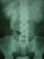
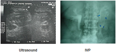
 A 47-year-old gentleman presented history of dull pain in the right flank of 6-months duration. Ultrasonography showed evidence of multiple gallstones and a thick walled gall bladder and gross hydronephrosis of the right kidney. IVU showed delayed excretion and drainage of the contrast and classical sea-horse sign (reverse J) confirming retrocaval ureter. Cystoscopy with retrograde urography confirmed our diagnosis. A Zebra wire was placed, which easily bypass the obstruction. A ureteric catheter was advanced to just below the obstruction. In the right lateral position with a 70 degree tilt, the same 4 ports were used. Right colon was mobilized adequately; ureter and inferior vena cava was exposed. The dilated upper ureter was mobilized till it started to wind around the inferior vena cava. The upper ureter was transacted and the circumcaval segment was transposed anteriorly. Uretero-ureteral anastomosis was performed with 4-0 vicryl. There was same difficulty in placing a stent. Therefore ureteric catheter was placed over the Zebra wire in position. The operating time was 240 minutes. Blood loss was insignificant. Patient made speedy recovery and resumed normal activity after 10 days. At 3-months follow-up an IVU showed good drainage.

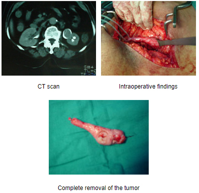
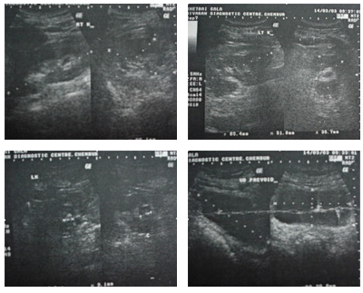
A 58-year-old lady had history of chronic left loin pain associated with repeated episodes of urinary tract infection. Ultrasonography showed ill-defined hypoechoic mass extending from renal pelvis to the upper ureter. IVU showed a filling defect extending from inferior group of calyces upto the upper ureter on the left side. CT scan confirmed a soft tissue lesion within the lumen of the left pelvis and upper ureter with an irregular hyperdense area seen within. Cystoscopy with left sided RGP confirmed a huge filling defect in the upper ureter and the pelvis. Diagnostic ureterorenoscopy was performed and a smooth globular large whitish tumour was seen extending from the upper ureter at L-3 and extending into the pelvis. Ureteroscopic biopsies showed evidence of normal mucosa with no suspicion of malignancy. A left sub-costal incision was used to expose the left upper ureter and pelvis grossly bulging tumour was felt from the outside. A U-shaped uretero-pyelotomy was performed and the entire lesion extending from the upper ureter to the lower calyx was delivered out of the incision. There were three vascular fronds attached to the mucosa, which were separately diathermised. Frozen section of the tumour showed no evidence of malignancy, the mass was suspected to be a benign fibroepithelial polyp. The lady had an uneventful post-operative stay and was home in 5 days. Her DJ stent was removed after 3 weeks. Histopathology confirmed a giant fibroepithelial polyp.

INVESTIGATIONS
Urine
- Pus Cell -15 - 20 / hpf
- RBC -Absent
URINE CULTURE : Sterile
S. BIOCHEMISTRY : Normal
USG

Right kidney : Normal
Left kidney :
- Lobulated outline
- Dilated PC system
- Echogenic area with distal shadowing
- seen in the inferior group of calyces
- Thickening of peri-renal tissue
